Thursday, October 30, 2014

The vestibular system and vision

I've been reading up on the link between vision and the vestibular system. It's been quite instructive to say the least. Of course it's annoying not to be able to align the eyes and have double vision while reading etc... However, there's more. It's very interesting how compromised eye movements, in large part due to eye muscle surgery, and consequently a compromised Vestibulo-Ocular Reflex are likely to be the biggest source of disability and fatigue for me personally. When going beyond my relatively low visual endurance limit, the sensory conflict between the visual and the vestibular gets exacerbated. It sometimes feels like being drunk and can even give me something resembling to a hangover later on. Not merely alignment of the eyes but also the integration of these ocular motor skills with the vestibular system will be crucial in terms of lasting vision therapy results and daily functionality.

What is the vestibular system?

- Peripheral
The term 'vestibular' comes from the Latin 'vestibule' which means room. This 'room' is located in the inner ear and contains a minute amount of chemical fluid. Its workings involve the detection of fluid movement by receptor hair cells in various differently oriented semicircular canals. Vestibular injury may or may not be accompanied by hearing loss.


- Central
The vestibular sense is often misunderstood or overlooked. That's because, for instance, there is no such thing as a primary vestibular cortex.

'There is, rather, a complex of cortical targets many of which remain difficult to understand. From the brainstem, vestibular signals reach a subcortical structure called the diencephalon. Tucked in between the cerebral hemispheres the diencephalon houses such important structures as the pineal gland, hypothalamus, and thalamus. Compared to the cerebral cortex the diencephalon is small in size but is very rich in nuclei for sensory, motor and limbic pathways. The thalamus is a central relay station for all incoming sensory signals excepting olfactory information.' - Vision Rehabilitation

Without going into detail, we can conclude this sense is for a large part located in the primitive,  reptilian part of our brain which houses many basic but vital functions. In fact, this sense is so essential to our functioning and well-being, that the vestibular nerve is the first fiber tract in the brain to begin myelination during pregnancy. This process starts by the last week of the first trimester. By the end of the fifth month of pregnancy, the vestibular organ has reached full size with ongoing myelination of pathways to the eyes and the spinal cord.

- Function
Whereas the anatomy and chemistry of the vestibular system itself are fascinating, its interaction with the visual system and other somato-sensory systems are even more interesting.

The vestibular system is responsible for
(1) detecting linear and angular head movement and head position in space;
(2) assisting gaze stabilization of the visual field;
(3) maintaining balance and postural control; 
(4) providing spatial orientation or perception of body movement.

The primary role of the vestibular system is to provide the brain with information to regulate posture and to coordinate eye and head movements. Proper vestibular functioning and integration with the other senses is of major importance to good overall health, optimal functionality and vision.

Link with vision?

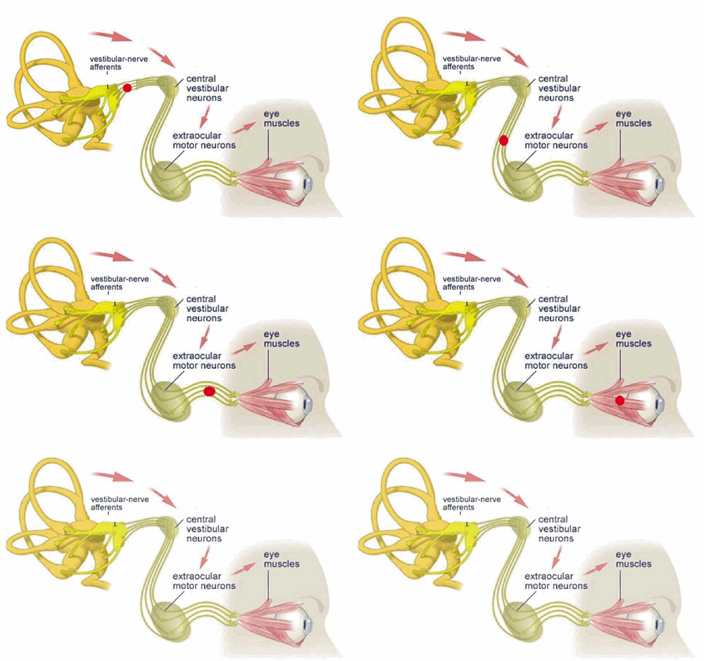
'The vestibular system plays an important role in the generation of eye movements that compensate for head movements. Through vestibular nuclei in the brainstem, each SCC (semi-circular canal) is able to communicate with motor neurons of extra-ocular muscles to cause eye deviation in each canal’s own plane. This interaction is the basis of the vestibuloocular reflex (VOR), which stabilizes gaze upon an image or the visual world during head movement. If the VOR is impaired, loss of gaze stabilization is the result. For example, as the patient turns the head while walking an object in the visual field or the whole visual environment appears unsteady or “bouncing.”

Demer described the VOR as a “synergistic” interaction between the vestibular and ocular systems. Normal VOR stabilization is needed for “functional vision, and vision optimizes the performance of the VOR”. Dynamic visual acuity (DVA) plays a role in maintaining a sharper image on the retina, while the VOR steadies gaze during head movement. This interaction is called the visual vestibuloocular reflex (VVOR).' - Vision Rehabilitation


And in turn...

'Sensory receptors in the eyes provide important feedback regarding orientation of the body in space. In other words, where is the head in relation to the horizon? As discussed in Chapters 5 through 7, dysfunction in the visual perception of “straight ahead,” common following brain injury, can cause dizziness, disorientation, and imbalance. In the process of orientation, visual input also provides information regarding possible environmental hazards, barriers or avenues of escape. In the presence of impaired inputs from either the somatosensory (sensory projections from all over the body) or vestibular or both systems there is a strong dependence upon visual input. Defective input from one of the three systems (visual, proprioception/somatosensory, vestibular) may be compensated for by the remaining intact systems such that the patient is able to function, albeit with some discomfort and loss of efficiency. However, dysfunction in two of the three systems will result in significant disability.' - Vision Rehabilitation

Problems?

Even though the vestibular sense might often be overlooked by physicians, one will be made aware of its existence in case of dysfunction.

- Inappropriate nystagmus
'Spontaneous nystagmus, occurring when the head is static (still), is an indicator of acute peripheral vestibular loss. Nystagmus is often along the horizontal plane. Vertical or torsional spontaneous nystagmus usually indicates a direct injury to the brainstem. The cause of this inappropriate nystagmus is a direction-specific imbalance in the VOR as brainstem neuronal circuitry is activated. Inappropriate nystagmus may also be related to medication toxicity, such as some antiseizure medications.' - Vision Rehabilitation

Inappropriate nystagmus may also be provoked by head shaking and indicates an imbalance of dynamic vestibular function.

An indicator of inappropriate nystagmus of central origin, caused by brain dysfunction rather than dysfunction of the peripheral organ, is often the inability to inhibit nystagmus with visual fixation.


- Dizziness
Feeling disoriented, especially in dark areas, or feeling spacey, floating, rocking, or lightheaded. Dizziness is not always related to vestibular dysfunction but may indicate non-vestibular causes, one of which being visual disorders. However, when dizziness is mentioned, involvement of a vestibular dysfunction must be considered.

- Vertigo
Even though dizziness and vertigo are often used interchangeably, technically there is a difference. Vertigo refers to the illusion of movement, typically rotational, when no actual movement is observed. True vertigo refers to vestibular system involvement.

- Motion sickness
What causes motion sickness?

'There is a hierarchical preference to rely on visual input more than any one of the other two systems (vestibular, proprioception) involved in balance. Vision, as proposed by Bronstein, had a dominant role or “hierarchical preference” over proprioceptive input in the process of maintaining upright posture and balance. However, Mallinson et al. suggested that a “visual preference strategy” would not apply to some individuals who have an intolerance for any disagreement between visual and vestibular signals. Paige had previously called this disagreement between the two systems as a “visual vestibular mismatch” (VVM) and others referred to it as a “sensory conflict.” By 1996, the “sensory conflict” theory for the development of motion sickness had been accepted within the scientific field studying this subject.' - Vision Rehabilitation

- And more...
Balance problems, gait ataxia, ...

Of the top of my head I can think of two interesting cases I read about involving vestibular dysfunction.

In the book 'The brain that changes itself', Norman Doidge described a woman who felt like she was perpetually falling due to vestibular injury.   Her vestibular organ was damaged but her central vestibular processing capabilities were intact. We can't even start to imagine how she felt. It completely ruined her life. Until a certain Doctor thought of the idea to supply her brain with 'vestibular input' in a different way. He connected an accelerometer to a tongue outlet which gave her a certain signal depending on the direction towards she was falling. Through the mechanisms of brain plasticity she learned to ignore her vestibular organ and to stabilize herself using this new input stream. Somehow over time, as her brain re-calibrated, she became less dependent on the device and needed it less and less.

Another vestibular story was featured in Oliver Sacks' 'The man who mistook his wife for a hat'. A Parkinsonian, 93 year old, retired carpenter whose vestibular sense had become more and more unreliable started walking tilted over to one side without knowing it. His senses deceived him until he saw himself on camera. He was speechless. After this discovery he wondered whether he could apply the mechanism of a spirit level to his problem. He designed a pair of glasses including some sort of spirit level which he could then use to teach himself to walk upright again. Essentially he retrained his impaired vestibular system using visual feedback. At first it took a lot of conscious effort but later on it got easier and easier. Just like learning how to drive a car.

Psychological impact of vestibular dysfunction

'There are a variety of reasons why patients suffering from episodes of vertigo develop anxiety, depression, poor self-esteem, and a growing sense of disability. The fear of falling or the fear of heights is common. Concern for a sudden onset of vertigo and the loss of control in public venues often leads to avoidance behaviors or “safety-seeking behaviors,” as coined by Gurr and Moffat. The appearance of staggering, falling, and confusion may lead observers to believe that the person is intoxicated. The patient soon finds him/herself avoiding social events and staying home to avoid embarrassment. Physical activity is avoided for fear of provoking an episode.' - Vision Rehabilitation


Testing

- Static and dynamic balance testing. Dynamic balance tests require input from the Vestibulo-Ocular Reflex to stabilize the visual field and make rapid adjustments in posture. Dynamic gait, sit-to-stand, ...
Dix Hallpike maneuver
- Bithermal Caloric irrigation test (COWS): spraying water in the ear. Check it out on Youtube. It's pretty cool.
- Comprehensive Vision Testing: gaze, tracking, binocular vision, perceptual, functional, nystagmography, ...

Please read Chapter 9 of 'Vision Rehabilitation' for more detailed information. 

Rehabilitiation of the vestibular system

Vestibular injury or dysfunction can impose dramatic alterations in a patient’s once active lifestyle. There will be occasions when medication can be helpful in the short term. Surgery is the rare choice. Therapists take caution when a patient has other disabilities (brain trauma, stroke, peripheral neuropathy, spinal conditions, and/or vision disorders), which may limit choices of exercises.

'A key factor in achieving the best outcome will be patient compliance. Education is the greatest tool used by a good therapist to engage a patient’s willing participation in what is usually a difficult treatment program. Between, both, the vestibular therapist and the psychotherapist the patient is assisted in understanding the basics of the dysfunction: Why symptoms occur; the emotional response; and how treatment should help. Initial treatment typically provokes unpleasant symptoms but feeling worse comes before feeling better. Another factor in achieving the best outcome involves adequate intensity and duration of the treatment program.

Progression of function is the best gauge for the effectiveness of treatment. The process of treatment involves strategies for adaptation and substitution by using other strategies, habituation (desensitization), and retraining of balance. As the patient is able, exercises are expanded to include cardiorespiratory conditioning. Goals of vestibular rehabilitation are to (1) optimize compensation in the balance system; (2) habituate abnormal vestibular responses to rapid movements; (3) reduce fall risks by improving balance and postural control; and (4) educate the patient.' - Vision Rehabilitation

Is it a vision problem or vestibular problem?

It's important to figure out whether you are suffering from a vestibular problem with a visual component or a vision problem with a vestibular component. For strabismics who identify with some of these issues, the answer is probably 'a vision problem with a vestibular component'.

That is why in the more advanced stages of Vision Therapy you are not only training eye movement. You are to stimulate your way towards overall sensory and thus vestibulo-visual integration. There's a million ways to skin that cat. Some of the things I do as vision therapy loading: walking, head turns while attempting to keep my gaze stable, walking a beam with yoked prisms, balance board, jumping on a trampoline, standing on my head, running, ...

When my vision tends to go blurry or double and I feel dizzy, it's time to take a break. I flirt with my limit, take sufficient rest and remind myself that sometimes you do have to feel worse before feeling better. Improving these elementary vestibulo-ocular skills does influence and improve more sophisticated and more cognitively taxing ocular motor activities such as reading. The ground is being prepared!

Thursday, October 9, 2014

'The ticking time bomb that split my world in two'

About a week ago me and a man named Ryan Brooks had a thought-provoking conversation on my Facebook wall.  I had just shared my latest blog entry 'The margin of error'. He commented "I always find your posts extremely interesting. I had a bleed in the brain stem which has left me with double vision. This is how I see now."


That is not your every day Facebook comment but I knew the visual phenomenon he was talking about of course. As I'm always open to learning about how other people deal with visual brain problems, I asked him whether he wanted to share more about what happened to him. He was prepared to do so and I am happy for it. Even though our histories are different (developmental vs cerebral incident), we are talking about closely related visual issues and symptoms. Thank you for sharing your incredible story, Ryan!

I present to you, Mr. Ryan Brooks.

Part 1
RYAN: "I am 40 years old. I live in Newcastle NSW, Australia. I was an occupational health and safety manager in the mining industry and traveled the world to places like New Caledonia and New Guinea to name a few. I had only been home for eight weeks when the incident happened. Over a period of three months prior to the bleed I had experienced three separate dizzy spells and racing heart episodes but only lasted for about ten minutes. I attributed it to maybe anxiety due to my stressful job.

The brain bleed happened on the 25th of June, 2012 at 11.15AM, I woke up feeling great and drove down to a cafe on the beach for a coffee. As I walked down the beach to the cafe my vision started to blur and I felt a strange buzzing feeling at the back of my head near the base of the skull. I put it down to still being half asleep. I ordered a coffee and sat at a table outside watching the surf. The buzzing started to get worse. I started to feel like I had butterflies in my stomach and felt increasingly faint. I got up to go to the bathroom to wash my face. As I got to the door... Boom. Suddenly I felt as if pins and needles were penetrating my face. Down my left arm and leg I felt numbness as well as pins and needles. All this combined with a feeling of complete disorientation. I managed to stumble back to the front counter and said "Call an ambulance, I'm having a stroke." Then I passed out over a table. A short while after, I came back to and noticed no one was helping me. I was in a bad way. I remembered there was no phone signal inside so I got on my hands and knees and crawled out the front door. I laid on the path, rang my sister and told her what had happened and for her to call an ambulance. The people at the bar told my sister that they thought I was a drug addict having an overdose. My sister went absolutely ballistic. The ambulance arrived 20 minutes later and off to hospital we went. After some brain scans the doctors realized that I had a bleed in the upper mid brain, which is part of the brain stem. More conspicuously, by merely looking at me, they could see my left eye was turned in so hard towards my nose you could barely see it. "


Ryan and his daughter


 Part 2
"The official diagnosis was a cavernous malformation. In plain English this means that a blood vessel wasn't formed properly at birth and it took 38 years to wear out and rupture. In other words, a ticking time bomb."

MICHAEL: After the incident and the diagnosis, did they do something about that blood vessel?

RYAN: "They did an angiogram and injected ink into my brain which basically gave them a map of all the blood vessels in my brain and of the problematic area of vessels in particular. They didn't do anything because the brain area where it happened is very complex. The vessels in that area of the brain stem are the size of a strand of hair. Fortunately they were not arteries. Doing something to 'fix' it would entail too much risk. An intervention might kill me or give me a major stroke. Instead we opted for yearly brain scans. There's no guarantee it won't happen again."

MICHAEL: How did things proceed from there on out?

RYAN: "After four initial weeks of rehab for partial paralysis down my left side everything started to get back to normal. Sort of... Concerns regarding double vision as a result of the bleed and nerve damage remained.

MICHAEL: Can you describe your current visual situation?

RYAN: "The official definition of the visual dysfunctions caused by the brain bleed goes as follows. Bilateral asymmetrical superior oblique paresis, cranial nerve paresis with convergence retraction nystagmus and some myopia in the left eye. But I would like to try and explain what that really means from my point of view.

From the outside my eye alignment looks normal other than a slight turn of the left eye.  Yet from the inside that's a different story. When I look straight ahead the image of my right eye looks pretty level but the image of my left eye is on a 45 degree vertical angle.The left eye also has very bad torsion.


When I tilt my head up and down the torsion becomes worse.  I have vertical up gaze palsy and so when I try to look up my eyes wiggle. Notwithstanding, when I tilt my head slightly to the right it pulls the torsion nearly square. In that moment my brain relaxes because it almost feels like I have normal and steady binocular vision again. Sadly I can't sustain the binocular posture. It's like a stare and after five seconds my left eye starts to drift.


When I turn my head left and right the images split up and down so I have to patch my left eye. It is very hard after 40 years of normal sight to now have two separate images that 'do what they do'.


When I walk without the left eye covered, the image coming from that eye bounces. So when using my right eye, with my left bad eye covered, the eye's movement compensates for any head movement. The image stays stable and straight (proper Vestibulo-Ocular Reflex). On the other hand, when I close my good right eye and only have to draw on my left eye, the image inadvertently moves up and down following head movement (compromised Vestibulo-Ocular Reflex). Not only does the image move up and down, but it also tilts because of the torsion. It's quite nauseating and throws my balance out. If I walk with only my bad left eye open,  it feels like I'm walking across a very steep hill. The image just bounces around inconsistently.

I have also noticed that the color isn't as good in my good right eye. At night my right good eye doesn't see as well as my left bad eye. Everything is a sort of tinted darker when looking through my right eye.

When I close one eye, my vision feels quite normal. although I had to get used to the difference in depth perception. At night, when there is little light, it feels like my vision is back to normal which stops the headaches. That's the only time my brain isn't fighting to get properly aligned binocular input. Then I can relax.

Considering all the above, using my vision is a very fine balancing act."

MICHAEL: How do you make these example images? Do you use an app?

RYAN: "You are actually the one who inspired me to investigate the internet and thus coming across the apps to help explain what I see to other people. These are the two apps. My Doctor was very impressed with the idea of taking photos of what I see and simulate what I see as I move my head into different directions. The images shift when doing so. This way he can see exactly what I see."


MICHAEL: What kind of treatment have you been getting or participating in?

RYAN: "I have been seeing a strabismus specialist in Melbourne at the Private Eye Clinic by the name of Dr Lional Kowal. I had VT for 12 months. While doing VT I was also going to Melbourne every three months for examinations. The VT pulled my eye nearly back to its normal position but the double vision remained.

After that initial year of VT, I have had five correctional surgeries but none have been successful due to damage to the 3rd, 4th, 6th, 9th and 10th cranial nerve. Every time a surgery was done it had a negative effect on either the vertical or horizontal relative positioning of the images. Unfortunately after five surgeries, two of which were to undo previously performed surgeries which had made my double vision worse, I am back to square one."

MICHAEL: I'm just thinking out loud... You had the incident in June 2012. Then you had one year of Vision Therapy aka Visual Neurorehabilitation. That brings us up to August 2013. So you must have had five surgeries in one year? How did that go? You seemed to be making VT progress, albeit slow. Who proposed the surgery? What did the Doctor tell you about its effectiveness, risks and possible outcomes?

RYAN: "As the double vision wasn't disappearing mainly due to the torsion aspect of my case, the Doctor or Neuro-Ophthalmologist suggested surgery. They explained the risks. He was confident he could give me an improvement.

The first operation was performed on both eyes and made the double vision worse. Five days later that surgery had to be undone.

Another three months later I had more eye muscle surgery on both eyes while being awake using only the use of anesthetic drops. They placed a black letter T on the ceiling and also on the wall in front of me. He started with the left eye. Cutting and manoeuvring the muscle a millimeter at a time. Then he'd wash out the eye, take the eye clamps out and tell me to look at the T and ask if there was any difference. Then he'd sit me up and tell me to look at the T on the wall in front of me and ask whether there was any difference. My response was 'it has adjusted slightly'. So back down for more adjusting. This process went on thee times until he said he couldn't do any more. He stated that if he overstretched the muscle it was irreversible. Next we repeated the same process for the right eye.


It was an extremely stressful experience. I could hear my heart racing. I was boiling hot due to the lights and surgical blankets. To be honest, after you have had your eye ball cut open, the eye muscles cut  and then flushed out with water, the mix of blood and water makes your vision quite blurry. So any indication of what I was seeing was compromised and not really accurate. My eyes kept drifting as my muscles were cut, my vision was blurred and the environment was extremely stressful.

A week later after letting my eyes recover a bit the double vision was worse. Another week later I went in for surgery again in an attempt to undo things. Under total anesthesia this time.

Four months later, one more surgery was done to do some slight tweaking. I was completely under for this one. Whatever the result was, I was going to have to live with it."

MICHAEL: You said 'I am back to square one'. Do you feel the surgeries were just a useless exercise or would you say it deteriorated the situation? I'm asking out of genuine curiosity, not because I myself had a very bad experience with strabismus surgery.

RYAN: "Now it is more or less back to where it was after the brain bleed, although a little different. Looking back I'm glad we gave it a try because now I know that at least we tried. I put my trust in my Doctor and we came out the other side not having gained but not having lost much either. He was a wonderful Doctor and only charged me for the first surgery. All the other surgeries he put through my medical fund at a significant price reduction. That was an incredibly kind gesture and showed that he was more interested in my case than in my money. On top of that I was extremely fortunate to have wonderful friends and family who organized a charity events such as a dinner party, an auction and a golf day. They ended up raising enough money to cover my surgeries, anesthetist, flights, accommodation, check ups, glasses, lenses, ... That all amounted to 30.000 dollars. If they read this, I want to thank Brett & Gail Purcell, Nathan Palmer, Tim & Macushla Spencer, Chad Edwards, Leeanne & Jeremy Symes and many other friends and family members who were involved. I had been 14 months off work so without this bunch of phenomenal friends my treatment would definitely have been cut short. I'm immensely grateful to them all."


Part 3
"Nowadays I'm using a tailor made contact lens that looks normal but really isn't. The outer edges are clear so you can still see the colour of my eye but the area covering my pupil is completely blacked out. I had to have my pupil measured so it looks cohesive with my other eye. This means that I am basically blind in my left eye when I have the lens in. Well, not completely blind... I still get peripheral vision from my left eye as the dot on the lens is a little smaller than my pupil and pupil size is variable depending on light conditions. Therefore, I still wear my glasses.   When I'm not wearing my glasses, I have learnt to ghost the images of my left eye but after a while this gives me head aches. So back on go the glasses! Looking at me you couldn't tell anything is wrong. However, without my blacked out lens and my glasses the double vision still gives me constant headaches. .

In general, my life has changed considerably following my accident. I no longer work in the mining industry. I now work as a disability case manager and spend much more time at home with my beautiful daughter."